Conditions treated
Our experts provide care for various conditions, including care for:
-
Achilles tendon rupture
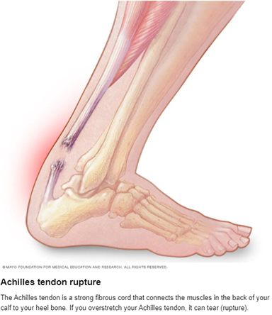
Achilles (uh-KILL-eez) tendon rupture is an injury that affects the back of your lower leg. It mainly occurs in people playing recreational sports, but it can happen to anyone.
The Achilles tendon is a strong fibrous cord that connects the muscles in the back of your calf to your heel bone. If you overstretch your Achilles tendon, it can tear (rupture) completely or just partially.
If your Achilles tendon ruptures, you might hear a pop, followed by an immediate sharp pain in the back of your ankle and lower leg that is likely to affect your ability to walk properly. Surgery is often performed to repair the rupture. For many people, however, nonsurgical treatment works just as well.
Symptoms
Although it's possible to have no signs or symptoms with an Achilles tendon rupture, most people have:
- The feeling of having been kicked in the calf
- Pain, possibly severe, and swelling near the heel
- An inability to bend the foot downward or "push off" the injured leg when walking
- An inability to stand on the toes on the injured leg
- A popping or snapping sound when the injury occurs
When to see your doctor
Seek medical advice immediately if you hear a pop in your heel, especially if you can't walk properly afterward.
Causes
Your Achilles tendon helps you point your foot downward, rise on your toes and push off your foot as you walk. You rely on it virtually every time you walk and move your foot.
Rupture usually occurs in the section of the tendon situated within 2 1/2 inches (about 6 centimeters) of the point where it attaches to the heel bone. This section might be prone to rupture because blood flow is poor, which also can impair its ability to heal.
Ruptures often are caused by a sudden increase in the stress on your Achilles tendon. Common examples include:
- Increasing the intensity of sports participation, especially in sports that involve jumping
- Falling from a height
- Stepping into a hole
Risk factors
Factors that may increase your risk of Achilles tendon rupture include:
- Age. The peak age for Achilles tendon rupture is 30 to 40.
- Sex. Achilles tendon rupture is up to five times more likely to occur in men than in women.
- Recreational sports. Achilles tendon injuries occur more often during sports that involve running, jumping, and sudden starts and stops — such as soccer, basketball and tennis.
- Steroid injections. Doctors sometimes inject steroids into an ankle joint to reduce pain and inflammation. However, this medication can weaken nearby tendons and has been associated with Achilles tendon ruptures.
- Certain antibiotics. Fluoroquinolone antibiotics, such as ciprofloxacin (Cipro) or levofloxacin (Levaquin), increase the risk of Achilles tendon rupture.
- Obesity. Excess weight puts more strain on the tendon.
Prevention
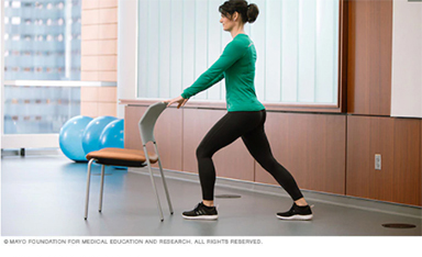
To reduce your chance of developing Achilles tendon problems, follow these tips:
- Stretch and strengthen calf muscles. Stretch your calf until you feel a noticeable pull but not pain. Don't bounce during a stretch. Calf-strengthening exercises can also help the muscle and tendon absorb more force and prevent injury. To do the stretch, follow these steps:
- Stand at arm's length from a wall or a sturdy piece of exercise equipment. Put your palms flat against the wall or hold on to the piece of equipment.
- Keep one leg back with your knee straight and your heel flat on the floor.
- Slowly bend your elbows and front knee and move your hips forward until you feel a stretch in your calf.
- Hold this position for 30 to 60 seconds.
- Switch leg positions and repeat with your other leg.
- Vary your exercises. Alternate high-impact sports, such as running, with low-impact sports, such as walking, biking or swimming. Avoid activities that place excessive stress on your Achilles tendons, such as hill running and jumping activities.
- Choose running surfaces carefully. Avoid or limit running on hard or slippery surfaces. Dress properly for cold-weather training, and wear well-fitting athletic shoes with proper cushioning in the heels.
- Increase training intensity slowly. Achilles tendon injuries commonly occur after an abrupt increase in training intensity. Increase the distance, duration and frequency of your training by no more than 10 percent weekly.
Diagnosis
During the physical exam, your doctor will inspect your lower leg for tenderness and swelling. Your doctor might be able to feel a gap in your tendon if it has ruptured completely.
The doctor might ask you to kneel on a chair or lie on your stomach with your feet hanging over the end of the exam table. He or she might then squeeze your calf muscle to see if your foot will automatically flex. If it doesn't, you probably have ruptured your Achilles tendon.
If there's a question about the extent of your Achilles tendon injury — whether it's completely or only partially ruptured — your doctor might order an ultrasound or MRI scan. These painless procedures create images of the tissues of your body.
Treatment
Treatment for a ruptured Achilles tendon often depends on your age, activity level and the severity of your injury. In general, younger and more active people, particularly athletes, tend to choose surgery to repair a completely ruptured Achilles tendon, while older people are more likely to opt for nonsurgical treatment.
Recent studies, however, have shown fairly equal effectiveness of both surgical and nonsurgical management.
Nonsurgical treatment
This approach typically involves:
- Resting the tendon by using crutches
- Applying ice to the area
- Taking over-the-counter pain relievers
- Keeping the ankle from moving for the first few weeks, usually with a walking boot with heel wedges or a cast, with the foot flexed down
Nonoperative treatment avoids the risks associated with surgery, such as infection.
However, a nonsurgical approach might increase your chances of re-rupture and recovery can take longer, although recent studies indicate favorable outcomes in people treated nonsurgically if they start rehabilitation with weight bearing early.
Surgery
The procedure generally involves making an incision in the back of your lower leg and stitching the torn tendon together. Depending on the condition of the torn tissue, the repair might be reinforced with other tendons.
Complications can include infection and nerve damage. Minimally invasive procedures reduce infection rates over those of open procedures.
Rehabilitation
After either treatment, you'll have physical therapy exercises to strengthen your leg muscles and Achilles tendon. Most people return to their former level of activity within four to six months. It's important to continue strength and stability training after that because some problems can persist for up to a year.
A type of rehabilitation known as functional rehabilitation also focuses on coordination of body parts and how you move. The purpose is to return you to your highest level of performance, as an athlete or in your everyday life.
One review study concluded that if you have access to functional rehabilitation, you might do just as well with nonsurgical treatment as with surgery. More study is needed.
Rehabilitation after either surgical or nonsurgical management is also trending toward moving earlier and progressing faster. Studies are ongoing in this area also.
-
ACL injury
An anterior cruciate ligament (ACL) injury occurs when the anterior cruciate ligament, one of the key ligaments that helps stabilize the knee, is stretched, sprained or torn. These injuries often happen during activities that involve sudden stopping, pivoting, jumping or landing.
When treatment may be considered
If an ACL injury is suspected, prompt medical evaluation is important. Treatment depends on the severity of the injury and may include rest, rehabilitation and physical therapy to restore strength and stability, or surgery followed by rehabilitation for more significant tears.
Signs and reasons to seek medical care
- You hear or feel a pop in your knee at the time of injury.
- Severe knee pain prevents you from continuing normal activity.
- Swelling develops quickly after the injury.
- You are unable to fully bend or straighten your knee.
- Your knee feels unstable or gives way when standing or walking.
Because the knee is a complex joint, early evaluation can help determine the extent of the injury and guide the most appropriate treatment plan.
-
Ankle arthritis
Psoriatic arthritis
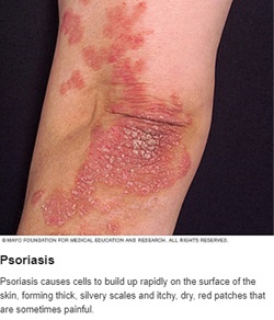
Psoriatic arthritis is a form of arthritis that affects some people who have psoriasis — a condition that features red patches of skin topped with silvery scales. Most people develop psoriasis first and are later diagnosed with psoriatic arthritis, but the joint problems can sometimes begin before skin patches appear.
Joint pain, stiffness and swelling are the main signs and symptoms of psoriatic arthritis. They can affect any part of your body, including your fingertips and spine, and can range from relatively mild to severe. In both psoriasis and psoriatic arthritis, disease flares may alternate with periods of remission.
No cure for psoriatic arthritis exists, so the focus is on controlling symptoms and preventing damage to your joints. Without treatment, psoriatic arthritis may be disabling.
Symptoms
Both psoriatic arthritis and psoriasis are chronic diseases that get worse over time, but you may have periods when your symptoms improve or go into remission alternating with times when symptoms become worse.
Psoriatic arthritis can affect joints on just one side or on both sides of your body. The signs and symptoms of psoriatic arthritis often resemble those of rheumatoid arthritis. Both diseases cause joints to become painful, swollen and warm to the touch.
However, psoriatic arthritis is more likely to also cause:
- Swollen fingers and toes. Psoriatic arthritis can cause a painful, sausage-like swelling of your fingers and toes. You may also develop swelling and deformities in your hands and feet before having significant joint symptoms.
- Foot pain. Psoriatic arthritis can also cause pain at the points where tendons and ligaments attach to your bones — especially at the back of your heel (Achilles tendinitis) or in the sole of your foot (plantar fasciitis).
- Lower back pain. Some people develop a condition called spondylitis as a result of psoriatic arthritis. Spondylitis mainly causes inflammation of the joints between the vertebrae of your spine and in the joints between your spine and pelvis (sacroiliitis).
When to see a doctor
If you have psoriasis, be sure to tell your doctor if you develop joint pain. Psoriatic arthritis can severely damage your joints if left untreated.
Causes
Psoriatic arthritis occurs when your body's immune system begins to attack healthy cells and tissue. The abnormal immune response causes inflammation in your joints as well as overproduction of skin cells.
It's not entirely clear why the immune system attacks healthy tissue, but it seems likely that both genetic and environmental factors play a role. Many people with psoriatic arthritis have a family history of either psoriasis or psoriatic arthritis. Researchers have discovered certain genetic markers that appear to be associated with psoriatic arthritis.
Physical trauma or something in the environment — such as a viral or bacterial infection — may trigger psoriatic arthritis in people with an inherited tendency.
Risk factors
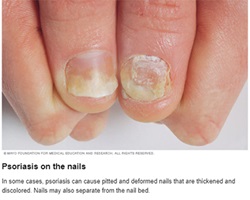
Several factors can increase your risk of psoriatic arthritis, including:
- Psoriasis. Having psoriasis is the single greatest risk factor for developing psoriatic arthritis. People who have pitted, deformed nails are especially likely to develop psoriatic arthritis.
- Your family history. Many people with psoriatic arthritis have a parent or a sibling with the disease.
- Your age. Although anyone can develop psoriatic arthritis, it occurs most often in adults between the ages of 30 and 50.
Complications
A small percentage of people with psoriatic arthritis develop arthritis mutilans — a severe, painful and disabling form of the disease. Over time, arthritis mutilans destroys the small bones in the hands, especially the fingers, leading to permanent deformity and disability.
People who have psoriatic arthritis sometimes also develop eye problems such as pinkeye (conjunctivitis) or uveitis, which can cause painful, reddened eyes and blurred vision. They are also at higher risk of cardiovascular disease.
Diagnosis
During the exam, your doctor may:
- Closely examine your joints for signs of swelling or tenderness
- Check your fingernails for pitting, flaking and other abnormalities
- Press on the soles of your feet and around your heels to find tender areas
No single test can confirm a diagnosis of psoriatic arthritis. But some types of tests can rule out other causes of joint pain, such as rheumatoid arthritis or gout.
Imaging tests
- X-rays. Plain X-rays can help pinpoint changes in the joints that occur in psoriatic arthritis but not in other arthritic conditions.
- Magnetic resonance imaging (MRI). MRI uses radio waves and a strong magnetic field to produce very detailed images of both hard and soft tissues in your body. This type of imaging test may be used to check for problems with the tendons and ligaments in your feet and lower back.
Laboratory tests
- Rheumatoid factor (RF). RF is an antibody that's often present in the blood of people with rheumatoid arthritis, but it's not usually in the blood of people with psoriatic arthritis. For that reason, this test can help your doctor distinguish between the two conditions.
- Joint fluid test. Using a needle, your doctor can remove a small sample of fluid from one of your affected joints — often the knee. Uric acid crystals in your joint fluid may indicate that you have gout rather than psoriatic arthritis.
Treatment
No cure exists for psoriatic arthritis, so treatment focuses on controlling inflammation in your affected joints to prevent joint pain and disability.
Medications
Drugs used to treat psoriatic arthritis include:
- NSAIDs. Nonsteroidal anti-inflammatory drugs (NSAIDs) can relieve pain and reduce inflammation. Over-the-counter NSAIDs include ibuprofen (Advil, Motrin IB, others) and naproxen sodium (Aleve). Stronger NSAIDs are available by prescription.
Side effects may include stomach irritation, heart problems, and liver and kidney damage. - Disease-modifying antirheumatic drugs (DMARDs). These drugs can slow the progression of psoriatic arthritis and save the joints and other tissues from permanent damage.
Common DMARDs include methotrexate (Trexall, Otrexup, others), leflunomide (Arava) and sulfasalazine (Azulfidine). Side effects vary but may include liver damage, bone marrow suppression and severe lung infections. - Immunosuppressants. These medications act to tame your immune system, which is out of control in psoriatic arthritis.
Examples include azathioprine (Imuran, Azasan) and cyclosporine (Gengraf, Neoral, Sandimmune). These medications can increase your susceptibility to infection. - Biologic agents. Also known as biologic response modifiers, this newer class of DMARDs includes abatacept (Orencia), adalimumab (Humira), certolizumab (Cimzia), etanercept (Enbrel), golimumab (Simponi), infliximab (Remicade), ixekizumab (Taltz), secukinumab (Cosentyx), tofacitinib (Xeljanz) and ustekinumab (Stelara).
These medications target specific parts of the immune system that trigger inflammation and lead to joint damage. These drugs can increase the risk of infections. Higher doses of tofacitinib can increase the risk of blood clots in the lungs. Biologic agents can be used alone or combined with DMARDs, such as methotrexate. - Newer oral medication. Apremilast (Otezla) decreases the activity of an enzyme in the body that controls the activity of inflammation within cells. Potential side effects include diarrhea, nausea and headaches.
Surgical and other procedures
- Steroid injections. This type of medication reduces inflammation quickly and is sometimes injected into an affected joint.
- Joint replacement surgery. Joints that have been severely damaged by psoriatic arthritis can be replaced with artificial prostheses made of metal and plastic.
Lifestyle and home remedies
- Protect your joints. Changing the way you carry out everyday tasks can make a tremendous difference in how you feel.
For example, you can avoid straining your finger joints by using gadgets such as jar openers to twist the lids from jars, by lifting heavy pans or other objects with both hands, and by pushing doors open with your whole body instead of just your fingers. - Maintain a healthy weight. Maintaining a healthy weight places less strain on your joints, leading to reduced pain and increased energy and mobility.
Losing weight, if needed, can also help your medications work better. Some psoriatic arthritis medications are less effective in people who have a body mass index of 25.0 to 29.9 (overweight).
The best way to increase nutrients while limiting calories is to eat more plant-based foods — fruits, vegetables and whole grains. - Exercise regularly. Exercise can help keep your joints flexible and your muscles strong. Types of exercises that are less stressful on joints include biking, swimming and walking.
- Stop smoking. Smoking is associated with a higher risk of developing psoriasis and with more-severe symptoms of psoriasis.
- Limit alcohol use. Alcohol can increase your risk of psoriasis, decrease the effectiveness of your treatment and increase side effects from some medications, such as methotrexate.
- Pace yourself. Battling pain and inflammation can leave you feeling exhausted. In addition, some arthritis medications can cause fatigue.
The key isn't to stop being active entirely, but to rest before you become too tired. Divide exercise or work activities into short segments. Find time to relax several times throughout the day.
Coping and support
Psoriatic arthritis can be particularly discouraging because the emotional pain that psoriasis can cause is compounded by joint pain and, in some cases, disability.
The support of friends and family can make a tremendous difference when you're facing the physical and psychological challenges of psoriatic arthritis. For some people, support groups can offer the same benefits.
A counselor or therapist can help you devise coping strategies to reduce your stress levels. The chemicals your body releases when you're under stress can aggravate both psoriasis and psoriatic arthritis.
Reactive arthritis
Reactive arthritis is joint pain and swelling triggered by an infection in another part of your body — most often your intestines, genitals or urinary tract.
Reactive arthritis usually targets your knees and the joints of your ankles and feet. Inflammation also can affect your eyes, skin and urethra.
Previously, reactive arthritis was sometimes called Reiter's syndrome, which was characterized by eye, urethra and joint inflammation.
Reactive arthritis isn't common. For most people, signs and symptoms come and go, eventually disappearing within 12 months.
Symptoms

The signs and symptoms of reactive arthritis generally start one to four weeks after exposure to a triggering infection. They might include:
- Pain and stiffness. The joint pain associated with reactive arthritis most commonly occurs in your knees, ankles and feet. You also might have pain in your heels, low back or buttocks.
- Eye inflammation. Many people who have reactive arthritis also develop eye inflammation (conjunctivitis).
- Urinary problems. Increased frequency and discomfort during urination may occur, as can inflammation of the prostate gland or cervix.
- Inflammation of soft tissue where it enters bone (enthesitis). This might include muscles, tendons and ligaments.
- Swollen toes or fingers. In some cases, your toes or fingers might become so swollen that they resemble sausages.
- Skin problems.Reactive arthritis can affect your skin a variety of ways, including a rash on your soles and palms and mouth sores.
- Low back pain. The pain tends to be worse at night or in the morning.
When to see a doctor
If you develop joint pain within a month of having diarrhea or a genital infection, contact your doctor.
Causes
Reactive arthritis develops in reaction to an infection in your body, often in your intestines, genitals or urinary tract. You might not be aware of the triggering infection if it causes mild symptoms or none at all.
Numerous bacteria can cause reactive arthritis. Some are transmitted sexually, and others are foodborne. The most common ones include:
- Chlamydia
- Salmonella
- Shigella
- Yersinia
- Campylobacter
- Clostridium difficile
Reactive arthritis isn't contagious. However, the bacteria that cause it can be transmitted sexually or in contaminated food. Only a few of the people who are exposed to these bacteria develop reactive arthritis.
Risk factors
Certain factors increase your risk of reactive arthritis:
- Age. Reactive arthritis occurs most frequently in adults between the ages of 20 and 40.
- Sex. Women and men are equally likely to develop reactive arthritis in response to foodborne infections. However, men are more likely than are women to develop reactive arthritis in response to sexually transmitted bacteria.
- Hereditary factors. A specific genetic marker has been linked to reactive arthritis. But many people who have this marker never develop the condition.
Prevention
Genetic factors appear to play a role in whether you're likely to develop reactive arthritis. Though you can't change your genetic makeup, you can reduce your exposure to the bacteria that may lead to reactive arthritis.
Make sure your food is stored at proper temperatures and is cooked properly to help you avoid the many foodborne bacteria that can cause reactive arthritis, including salmonella, shigella, yersinia and campylobacter. Some sexually transmitted infections can trigger reactive arthritis. Using condoms might lower your risk.
Diagnosis
During the physical exam, your doctor is likely to check your joints for signs and symptoms of inflammation, such as swelling, warmth and tenderness, and test range of motion in your spine and affected joints. Your doctor might also check your eyes for inflammation and your skin for rashes.
Blood tests
Your doctor might recommend that a sample of your blood be tested for:
- Evidence of past or current infection
- Signs of inflammation
- Antibodies associated with other types of arthritis
- A genetic marker linked to reactive arthritis
Joint fluid tests
Your doctor might use a needle to withdraw a sample of fluid from within an affected joint. This fluid will be tested for:
- White blood cell count. An increased number of white blood cells might indicate inflammation or an infection.
- Infections. Bacteria in your joint fluid might indicate septic arthritis, which can result in severe joint damage.
- Crystals. Uric acid crystals in your joint fluid might indicate gout. This very painful type of arthritis often affects the big toe.
Imaging tests
X-rays of your low back, pelvis and joints can indicate whether you have any of the characteristic signs of reactive arthritis.
Treatment
The goal of treatment is to manage your symptoms and treat an infection that could still be present.
Medications
If your reactive arthritis was triggered by a bacterial infection, your doctor might prescribe an antibiotic if there is evidence of persistent infection. Which antibiotic you take depends on the bacteria that are present.
Other medications might include:
- Nonsteroidal anti-inflammatory drugs (NSAIDs). Prescription NSAIDs, such as indomethacin (Indocin), can relieve the inflammation and pain of reactive arthritis.
- Corticosteroids. Injection of a corticosteroid into affected joints can reduce inflammation and allow you to return to your normal activity level.
- Topical steroids. These might be used for skin rashes caused by reactive arthritis.
- Rheumatoid arthritis drugs. Limited evidence suggests that medications such as sulfasalazine (Azulfidine), methotrexate (Trexall) or etanercept (Enbrel) can relieve pain and stiffness for some people with reactive arthritis.
Physical therapy
A physical therapist can provide you with targeted exercises for your joints and muscles. Strengthening exercises develop the muscles around your affected joints, which increase the joint's support. Range-of-motion exercises can increase your joints' flexibility and reduce stiffness.
Septic arthritis
Septic arthritis is a serious joint infection that can quickly damage cartilage and bone if not treated. The infection occurs when bacteria, viruses or fungi enter a joint, often through the bloodstream from another infection in the body. Septic arthritis most commonly affects the knee but also can occur in the hip, shoulder and other joints. Prompt diagnosis and treatment are important to help protect joint function and prevent lasting damage.
Symptoms of septic arthritis
Symptoms often develop suddenly and may include:
- Severe joint pain
- Joint swelling
- Redness or warmth around the joint
- Difficulty moving the joint
- Fever or chills
Seek medical care right away if you experience severe joint pain along with swelling, redness or fever.
Causes and risk factors
Septic arthritis develops when an infection spreads to a joint. Your risk may be higher if you:
- Have arthritis, gout, lupus or another joint condition.
- Have a weakened immune system.
- Have diabetes, kidney disease or liver disease.
- Have had a joint injury, surgery or joint replacement.
- Have a skin infection, open wound or puncture injury near a joint.
Diagnosis and treatment
To diagnose septic arthritis, your healthcare team may evaluate joint fluid, perform blood tests and order imaging studies. Treatment typically includes draining infected fluid from the joint and using antibiotics to clear the infection. Early treatment can help relieve symptoms, protect the joint and reduce the risk of permanent damage.
Potential complications
Without prompt treatment, septic arthritis can lead to permanent joint damage, loss of mobility and chronic joint problems.
-
Arthritis
Do you have swelling, pain and reduced motion in your hands and wrists? If you answer yes to this question, you may be suffering from arthritis. Our trained staff can help effectively manage your arthritis symptoms with medical and surgical therapies.
-
Broken foot

A broken foot is an injury to the bone. You may experience a broken foot during a car crash or from a simple misstep or fall.
The seriousness of a broken foot varies. Fractures can range from tiny cracks in your bones to breaks that pierce your skin.
Treatment for a broken foot depends on the exact site and severity of the fracture. A severely broken foot may require surgery to implant plates, rods or screws into the broken bone to maintain proper position during healing.
Symptoms
If you have a broken foot, you may experience some of the following signs and symptoms:
- Immediate, throbbing pain
- Pain increases with activity and decreases with rest
- Swelling
- Bruising
- Tenderness
- Deformity
- Difficulty in walking or bearing weight
When to see a doctor
See a doctor if there is obvious deformity, if the pain and swelling don't get better with self-care, or if the pain and swelling gets worse over time. Also, see a doctor if the injury interferes with walking.
Causes
The most common causes of a broken foot include:
- Car accidents. The crushing injuries common in car accidents may cause breaks that require surgical repair.
- Falls. Tripping and falling can break bones in your feet, as can landing on your feet after jumping down from just a slight height.
- Impact from a heavy weight. Dropping something heavy on your foot is a common cause of fractures.
- Missteps. Sometimes just putting your foot down wrong can result in a broken bone. A toe can get broken from stubbing your toes on furniture.
- Overuse. Stress fractures are common in the weight-bearing bones of your feet. These tiny cracks are usually caused over time by repetitive force or overuse, such as running long distances. But they can also occur with normal use of a bone that's been weakened by a condition such as osteoporosis.
Risk factors
You may be at higher risk of a broken foot or ankle if you:
- Participate in high-impact sports. The stresses, direct blows and twisting injuries that occur in sports such as basketball, football, gymnastics, tennis and soccer can causes foot fractures.
- Use improper technique or sports equipment. Faulty equipment, such as shoes that are too worn or not properly fitted, can contribute to stress fractures and falls. Improper training techniques, such as not warming up and stretching, also can cause foot injuries.
- Suddenly increase your activity level. Whether you're a trained athlete or someone who's just started exercising, suddenly boosting the frequency or duration of your exercise sessions can increase your risk of a stress fracture.
- Work in certain occupations. Certain work environments, such as a construction site, put you at risk of falling from a height or dropping something heavy on your foot.
- Keep your home cluttered or poorly lit. Walking around in a house with too much clutter or too little light may lead to falls and foot injuries.
- Have certain conditions. Having decreased bone density (osteoporosis) can put you at risk of injuries to your foot bones.
Complications
Complications of a broken foot are uncommon but may include:
- Arthritis. Fractures that extend into a joint can cause arthritis years later. If your foot starts to hurt long after a break, see your doctor for an evaluation.
- Bone infection (osteomyelitis). If you have an open fracture, meaning one end of the bone protrudes through the skin, your bone may be exposed to bacteria that cause infection.
- Nerve or blood vessel damage. Trauma to the foot can injure adjacent nerves and blood vessels, sometimes actually tearing them. Seek immediate attention if you notice any numbness or circulation problems. Lack of blood flow can cause a bone to die and collapse.
Prevention
These basic sports and safety tips may help prevent a broken foot:
- Wear proper shoes. Use hiking shoes on rough terrain. Wear steel-toed boots in your work environment if necessary. Choose appropriate athletic shoes for your sport.
- Replace athletic shoes regularly. Discard sneakers as soon as the tread or heel wears out or if the shoes are wearing unevenly. If you're a runner, replace your sneakers every 300 to 400 miles.
- Start slowly. That applies to a new fitness program and each individual workout.
- Cross-train. Alternating activities can prevent stress fractures. Rotate running with swimming or biking.
- Build bone strength. Calcium-rich foods, such as milk, yogurt and cheese, really can do your body good. Taking vitamin D supplements also can help.
- Use night lights. Many broken toes are the result of walking in the dark.
- Declutter your house. Keeping clutter off the floor can help you to avoid trips and falls.
Diagnosis
During the physical exam, your doctor will check for points of tenderness in your foot. The precise location of your pain can help determine its cause.
They may move your foot into different positions, to check your range of motion. You may be asked to walk for a short distance so that your doctor can examine your gait.
Imaging tests
If your signs and symptoms suggest a break or fracture, your doctor may suggest one or more of the following imaging tests.
- X-rays. Most foot fractures can be visualized on X-rays. The technician may need to take X-rays from several different angles so that the bone images won't overlap too much. Stress fractures often don't show up on X-rays until the break actually starts healing.
- Bone scan. For a bone scan, a technician will inject a small amount of radioactive material into a vein. The radioactive material is attracted to your bones, especially the parts of your bones that have been damaged. Damaged areas, including stress fractures, show up as bright spots on the resulting image.
- Computerized tomography (CT). CT scans take X-rays from many different angles and combine them to make cross-sectional images of internal structures of your body. CT scans can reveal more detail about the bone and the soft tissues that surround it, which may help your doctor determine the best treatment.
- Magnetic resonance imaging (MRI). MRI uses radio waves and a strong magnetic field to create very detailed images of the ligaments that help hold your foot and ankle together. This imaging helps to show ligaments and bones and can identify fractures not seen on X-rays.
Treatment
Treatments for a broken foot will vary, depending on which bone has been broken and the severity of the injury.
Medications
Your doctor may recommend an over-the-counter pain reliever, such as acetaminophen (Tylenol, others).
Therapy
After your bone has healed, you'll probably need to loosen up stiff muscles and ligaments in your feet. A physical therapist can teach you exercises to improve your flexibility and strength.
Surgical and other procedures
- Reduction. If you have a displaced fracture, meaning the two ends of the fracture are not aligned, your doctor may need to manipulate the pieces back into their proper positions — a process called reduction. Depending on the amount of pain and swelling you have, you may need a muscle relaxant, a sedative or even a general anesthetic before this procedure.
- Immobilization. To heal, a broken bone must be immobilized so that its ends can knit back together. In most cases, this requires a cast.
Minor foot fractures may only need a removable brace, boot or shoe with a stiff sole. A fractured toe is usually taped to a neighboring toe, with a piece of gauze between them. - Surgery. In some cases, an orthopedic surgeon may need to use pins, plates or screws to maintain proper position of your bones during healing. These materials may be removed after the fracture has healed if they are prominent or painful.
Preparing for your appointment
You will likely initially seek treatment for a broken foot in an emergency room or urgent care clinic. If the pieces of broken bone aren't lined up properly for healing, you may be referred to a doctor specializing in orthopedic surgery.
What you can do
You may want to write a list that includes:
- Detailed descriptions of your symptoms
- Information about medical problems you've had
- Information about the medical problems of your parents or siblings
- All the medications and dietary supplements you take
- Questions you want to ask the doctor
For a broken ankle or foot, basic questions to ask your doctor include:
- What tests are needed?
- What treatments are available, and which do you recommend?
- If I need a cast, how long will I need to wear it?
- Will I need surgery?
- What activity restrictions will need to be followed?
- Should I see a specialist?
- What pain medications do you recommend?
Don't hesitate to ask any other questions you have.
What to expect from your doctor
Your doctor may ask some of the following questions:
- Was there a specific injury that triggered your symptoms?
- Did your symptoms come on suddenly?
- Have you injured your feet in the past?
- Have you recently begun or intensified an exercise program?
What to do in the meantime
If your injury isn't severe enough to warrant a trip to the emergency room, here are some things you can do at home to care for your injury until you can see your doctor:
- Apply ice for 15 to 20 minutes at a time, every three to four hours to bring down the swelling.
- Keep your foot elevated.
- Don't put any weight on your injured foot.
- Lightly wrap the injury in a soft bandage that provides slight compression.
-
Bunions

A bunion is a bony bump that forms on the joint at the base of your big toe. It occurs when some of the bones in the front part of your foot move out of place. This causes the tip of your big toe to get pulled toward the smaller toes and forces the joint at the base of your big toe to stick out. The skin over the bunion might be red and sore.
Wearing tight, narrow shoes might cause bunions or make them worse. Bunions can also develop because of the shape of your foot, a foot deformity or a medical condition, such as arthritis.
Smaller bunions (bunionettes) can develop on the joint of your little toe.
Symptoms
The signs and symptoms of a bunion include:
- A bulging bump on the outside of the base of your big toe
- Swelling, redness or soreness around your big toe joint
- Corns or calluses — these often develop where the first and second toes rub against each other
- Ongoing pain or pain that comes and goes
- Limited movement of your big toe
When to see a doctor
Although bunions often require no medical treatment, see your doctor or a doctor who specializes in treating foot disorders (podiatrist or orthopedic foot specialist) if you have:
- Ongoing big toe or foot pain
- A visible bump on your big toe joint
- Decreased movement of your big toe or foot
- Difficulty finding shoes that fit properly because of a bunion
Causes
There are many theories about how bunions develop, but the exact cause is unknown. Factors likely include:
- Inherited foot type
- Foot stress or injuries
- Deformities present at birth
Experts disagree on whether tight, high-heeled or too-narrow shoes cause bunions or whether footwear simply contributes to the development of bunions.
Bunions might be associated with certain types of arthritis, particularly inflammatory types, such as rheumatoid arthritis.
Risk factors
These factors might increase your risk of bunions:
- High heels. Wearing high heels forces your toes into the front of your shoes, often crowding your toes.
- Ill-fitting shoes. People who wear shoes that are too tight, too narrow or too pointed are more likely to develop bunions.
- Rheumatoid arthritis. Having this inflammatory condition can make you more likely to develop bunions.
- Heredity. The tendency to develop bunions might be the result of an inherited problem with the structure or anatomy of your foot.
Complications
Possible complications of bunions include:
- Bursitis. This painful condition occurs when the small fluid-filled pads that cushion the bones near your joints become inflamed.
- Hammertoe. An abnormal bend that occurs in the middle joint of a toe, usually the toe next to your big toe, can cause pain and pressure.
- Metatarsalgia. This condition causes pain and swelling in the ball of your foot.
Prevention
To help prevent bunions, choose shoes carefully. They should have a wide toe box — no pointy toes — and there should be space between the tip of your longest toe and the end of the shoe.
Your shoes should conform to the shape of your feet without squeezing or pressing any part of your foot.
Diagnosis
Your doctor can identify a bunion by examining your foot. After the physical exam, an X-ray of your foot can help your doctor determine the best way to treat it.
Treatment
Treatment options vary depending on the severity of your bunion and how much pain it causes.
Conservative treatment
Nonsurgical treatments that may relieve the pain and pressure of a bunion include:
- Changing shoes. Wear roomy, comfortable shoes that provide plenty of space for your toes.
- Padding. Over the counter, nonmedicated bunion pads or cushions may be helpful. They can act as a buffer between your foot and your shoe and ease your pain.
- Medications. Acetaminophen (Tylenol, others), ibuprofen (Advil, Motrin IB, others) or naproxen sodium (Aleve) can help you control the pain of a bunion. Cortisone injections also might help. .
- Shoe inserts. Padded shoe inserts can help distribute pressure evenly when you move your feet, reducing your symptoms and preventing your bunion from getting worse. Over-the-counter supports can provide relief for some people; others require prescription orthotic devices.
- Applying ice. Icing your bunion after you've been on your feet too long or if it becomes inflamed can help relieve soreness and swelling. If you have reduced feeling or circulation problems with your feet, check with your doctor first before applying ice.
Surgical options
If conservative treatment doesn't relieve your symptoms, you might need surgery. Surgery is not recommended for cosmetic reasons; only when a bunion causes you frequent pain or interferes with your daily activities.
There are many surgical procedures for bunions, and no one technique is best for every problem.
Surgical procedures for bunions can be done as single procedures or in combination. They might involve:
- Removing the swollen tissue from around your big toe joint
- Straightening your big toe by removing part of the bone
- Realigning one or more bones in the forefoot to a more normal position to correct the abnormal angle in your big toe joint
- Joining the bones of your affected joint permanently
It's possible that you'll be able to walk on your foot right after a bunion procedure. However, full recovery can take weeks to months.
To prevent a recurrence, you'll need to wear proper shoes after recovery. For most people, it's unrealistic to expect to wear narrower shoes after surgery.
Talk to your doctor about what you can expect after bunion surgery.
-
Carpal tunnel syndrome
Using your hands and fingers is something you likely do without thinking, until numbness and tingling occur, which can prevent you from performing daily tasks. Carpal tunnel syndrome occurs when you have a pinched nerve in your wrist. If splinting and rest haven't helped, our experts will recommend surgery options to restore function in your hands and fingers.
Carpal tunnel anatomy
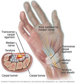
A passageway from the wrist to the hand, the carpal tunnel is made of tendons, ligaments and bones. The median nerve passes through the tunnel and provides sensation to your thumb, index finger, middle finger and the thumb-side of the ring finger.
Carpal tunnel release
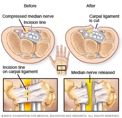
During carpal tunnel release, a surgeon makes an incision in the palm of your hand over the carpal tunnel ligament and cuts through the ligament to relieve pressure on the median nerve. The surgery may be done by making one incision on the palm side of the wrist, or by making several small incisions.
-
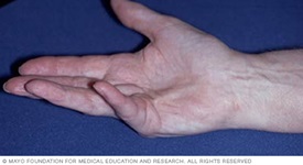
Dupuytren's contracture
Breaking your hand or wrist can happen easily during a fall, especially if your bones are thin and fragile from osteoporosis. It’s important to come see us right away if you have a fracture so that we can minimize your pain and get you back to normal function as quickly as possible.
-
Flatfeet
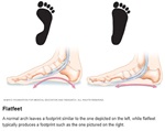
You have flatfeet when the arches on the inside of your feet are flattened, allowing the entire soles of your feet to touch the floor when you stand up.
A common and usually painless condition, flatfeet can occur when the arches don't develop during childhood. In other cases, flatfeet develop after an injury or from the simple wear-and-tear stresses of age.
Flatfeet can sometimes contribute to problems in your ankles and knees because the condition can alter the alignment of your legs. If you aren't having pain, no treatment is usually necessary for flatfeet.
Symptoms
Most people have no signs or symptoms associated with flatfeet. But some people with flatfeet experience foot pain, particularly in the heel or arch area. Pain may worsen with activity. Swelling along the inside of the ankle can also occur.
When to see a doctor
Talk to your doctor if you or your child has foot pain.
Causes
A flat foot is normal in infants and toddlers, because the foot's arch hasn't yet developed. Most people's arches develop throughout childhood, but some people never develop arches. This is a normal variation in foot type, and people without arches may or may not have problems.
Some children have flexible flatfoot, in which the arch is visible when the child is sitting or standing on tiptoes, but disappears when the child stands. Most children outgrow flexible flatfoot without problems.
Arches can also fall over time. Years of wear and tear can weaken the tendon that runs along the inside of your ankle and helps support your arch.
Risk factors
Factors that can increase your risk of flatfeet include:
- Obesity
- Injury to your foot or ankle
- Rheumatoid arthritis
- Aging
- Diabetes
Diagnosis
To view the mechanics of your feet, your doctor will observe your feet from the front and back and ask you to stand on your toes. He or she might also look at the wear pattern on your shoes.
Imaging tests
If you have a lot of pain in your feet, your doctor may order tests such as:
- X-rays. A simple X-ray uses a small amount of radiation to produce images of the bones and joints in your feet. It's particularly useful in detecting arthritis.
- CT scan. This test takes X-rays of your foot from different angles and provides much more detail than a standard X-ray.
- Ultrasound. If your doctor suspects an injured tendon, he or she may request this test, which uses sound waves to produce detailed images of soft tissues within the body.
- MRI. Using radio waves and a strong magnet, MRIs provide excellent detail of both hard and soft tissues.
Treatment
No treatment is necessary for flatfeet if they don't cause pain.
Therapy
If it is painful, your doctor might suggest:
- Arch supports (orthotic devices). Over-the-counter arch supports may help relieve the pain caused by flatfeet. Or your doctor might suggest custom-designed arch supports, which are molded to the contours of your feet. Arch supports won't cure flatfeet, but they often reduce symptoms.
- Stretching exercises. Some people with flatfeet also have a shortened Achilles tendon. Exercises to stretch this tendon may help.
- Supportive shoes. A structurally supportive shoe might be more comfortable than sandals or shoes with minimal support.
- Physical therapy. Flatfeet may contribute to overuse injuries in some runners. A physical therapist can do a video analysis of how you run to help you improve your form and technique.
Surgery
Surgery isn't done solely to correct flatfeet. However, you might have surgery for an associated problem, such as a tendon tear or rupture.
Lifestyle and home remedies
If your flatfeet cause minor pain, you might want to try:
- Rest. Avoid activities that aggravate your condition. Participate in low-impact activities — such as walking, biking or swimming — rather than jumping and running activities.
- Arch supports. Over-the-counter arch supports might increase your comfort.
- Medications. Over-the-counter pain relievers might help.
- Weight loss. Losing weight can reduce stress on your feet.
-
Hand and wrist pain and injury
Hand and wrist pain can make everyday activities difficult, from typing and cooking to lifting and gripping. Because the hand and wrist contain many bones, joints, tendons, ligaments and nerves in a small area, injuries and conditions affecting these structures can cause pain, swelling, weakness and loss of function. Orthopedic specialists diagnose and treat a wide range of hand and wrist concerns, helping patients find relief and restore movement.
When treatment may be considered
Evaluation by an orthopedic specialist may be recommended when hand or wrist symptoms are severe, continue despite rest and conservative care, or interfere with daily activities. Treatment may also be needed for injuries such as fractures, dislocations, tendon or ligament damage, or for chronic conditions that have not improved with nonsurgical approaches. In some cases, advanced treatments or surgery may be considered to relieve pain, improve function and address the underlying cause of the problem.
Signs you may need medical care
- Ongoing hand or wrist pain, swelling or stiffness that does not improve with rest or over-the-counter pain relievers.
- Injuries caused by a fall, sports activity or other trauma that may result in a fracture, dislocation, tendon injury or ligament damage.
- Weakness, reduced grip strength, numbness, tingling or difficulty moving the hand, fingers or wrist.
- Symptoms related to conditions such as arthritis, carpal tunnel syndrome, trigger finger or tendon inflammation that continue despite conservative treatment.
- Repetitive-use injuries from work, sports or daily activities that cause persistent pain, inflammation or reduced function.
-
Hip pain and injury
Mayo Clinic Health System orthopedic specialists diagnose and treat conditions that affect the hip and surrounding muscles, tendons, ligaments and joints. Whether hip pain is caused by an injury, arthritis or another condition, our team works with you to find the right treatment to reduce pain, improve mobility and help you return to daily activities.
When treatment may be considered
Evaluation and treatment may be appropriate when hip pain continues despite self-care measures such as rest, ice, heat or over-the-counter medications, or when symptoms begin to interfere with work, exercise, sleep or other everyday activities. Depending on the cause, treatment may include physical therapy, medications, injections or surgery. For some patients, procedures such as hip arthroscopy, hip preservation surgery or hip replacement may be considered when nonsurgical treatments are no longer providing relief.
Signs you may need orthopedic care for hip pain or injury
- Persistent hip pain that does not improve, worsens with activity, disrupts sleep or limits daily routines.
- Difficulty walking, standing, bearing weight on the affected leg or moving the hip normally.
- Hip stiffness, reduced range of motion or trouble performing everyday movements such as bending, sitting or getting up from a chair.
- Hip injuries or conditions including fractures, dislocations, labral tears, femoroacetabular impingement (FAI), osteoarthritis, rheumatoid arthritis, osteonecrosis or greater trochanteric pain syndrome.
- Ongoing symptoms that have not improved with conservative treatments such as physical therapy, activity modification or injections.
-
Knee pain and injury
Mayo Clinic Health System orthopedic specialists diagnose and treat conditions that affect the knee, including injuries, arthritis and other causes of pain, swelling and limited movement. Care is tailored to each person and may include nonsurgical treatments, rehabilitation or surgery when needed to help improve comfort, stability and function.
When treatment may be considered
It may be time to seek care if knee pain does not improve with rest, ice, activity changes or over-the-counter medications, or if symptoms are interfering with work, exercise or daily activities. In some cases, surgery may be recommended for significant injuries, advanced arthritis or when other treatments have not provided enough relief.
Reasons to seek care for knee pain or injury
- Ongoing pain or swelling that does not improve or is accompanied by redness, warmth or increasing discomfort.
- Knee instability or difficulty bearing weight, including a feeling that the knee is giving way or cannot support normal movement.
- Limited range of motion or visible changes in the shape or alignment of the knee or leg.
- Catching, locking, popping or grinding sensations that may signal cartilage, meniscus or kneecap-related problems.
- Arthritis-related symptoms, such as stiffness, pain and reduced mobility that affect everyday activities and quality of life.
-
Nerve lacerations
Your peripheral nerves are fragile and easily injured during trauma. If you had an injury that caused a laceration, you will feel numbness, weakness or tingling. If your nerve is lacerated or divided in two, our expert surgeons will reconnect the nerve ends.
-
Pediatric orthopedics
Your child, whether an infant or adolescent, may need specialized bone and joint care for orthopedic and developmental conditions. He or she will receive a personalized evaluation and treatment plan including nonsurgical and surgical treatments for:
- Scoliosis
- Spinal disorders
- Clubfoot
- Growth plate injuries
- Upper and lower extremity disorders
- Baby hip instability and dysplasia
- Pediatric and adolescent hip problems
- Cerebral Palsy
- Spina bifida
- Sports injuries
-
Shoulder pain and injury
Mayo Clinic Health System orthopedic specialists diagnose and treat conditions that affect the shoulder joint, muscles, tendons and ligaments. Whether shoulder pain develops gradually or occurs after an injury, our team works with you to identify the cause and recommend treatment options to help restore comfort, movement and function.
When Shoulder Treatment May Be Considered
Evaluation and treatment may be recommended when shoulder pain, weakness or limited movement does not improve with rest, ice, over-the-counter pain relief or other self-care measures. In some cases, surgery may be considered for significant injuries or advanced joint damage when conservative treatments have not provided enough relief.
Reasons to Seek Care for Shoulder Pain or Injury
- Ongoing or severe shoulder pain that interferes with daily activities, work, exercise or sleep.
- Limited range of motion, stiffness or weakness that makes it difficult to lift, reach or use your arm normally.
- Shoulder instability or deformity, including a feeling that the joint may slip out of place or visible changes after an injury.
- Common shoulder conditions, such as rotator cuff tears, frozen shoulder, shoulder dislocation, separated shoulder, tendinopathy or osteoarthritis.
- Symptoms that persist despite treatment, including pain or mobility issues that continue after physical therapy, activity modification or other nonsurgical approaches.
-
Tendinopathies
Inflammation of a tendon can occur in any part of your body, which causes tenderness and pain with any movement you make in that area. If you've tried rest, ice and over-the-counter pain relievers and are still experiencing pain, we can help. Our skilled providers will talk with you to decide if a steroid injection or surgery is best to relieve your pain and prevent further injury. Conditions that can result from tendonitis include trigger finger and De Quervain's tenosynovitis.
- Trigger finger — Trigger finger generally results from inflammation within a tendon sheath, restricting tendon motion. A bump (nodule) also may form.

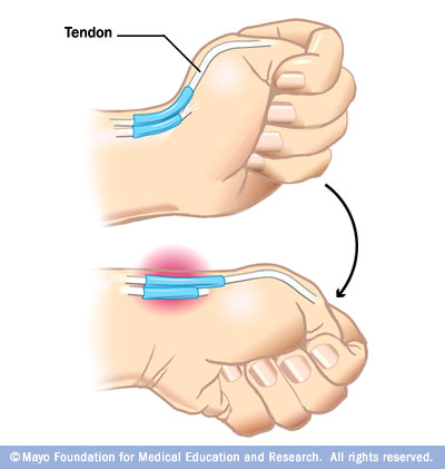
- De Quervain's tenosynovitis — A test called the Finkelstein test can help your doctor confirm de Quervain's tenosynovitis. To do this test, first bend your thumb down across the palm of your hand, covering your thumb with your fingers. Next, bend your wrist toward your little finger. If this causes pain, there is a chance you may have de Quervain's tenosynovitis.

Diagnosis & treatments
Orthopedic specialists provide diagnosis and surgical and nonsurgical treatment options that can help you enjoy an active, pain-free life. We've redesigned the orthopedic care experience, including a leading joint replacement program that allows you to recover more quickly and leave the hospital as soon as possible.
-
Ankle surgery
Ankle surgery helps relieve pain and improve mobility when nonsurgical treatments are no longer effective. At Mayo Clinic Health System, orthopedic specialists provide personalized care using proven surgical options to help you return to daily activities with greater comfort and stability.
When ankle surgery may be recommended
Surgery may be an option if ankle arthritis causes ongoing pain, stiffness, or limited movement despite treatments such as physical therapy, medications, activity changes, or supportive footwear and braces.
Your care team considers your symptoms, activity level, overall health, and how arthritis affects your quality of life before recommending surgery.
Understanding ankle arthritis
The ankle joint connects three bones — the tibia, fibula, and talus. Healthy cartilage allows smooth movement between these bones. Arthritis damages cartilage and joint surfaces, leading to pain, swelling, and reduced motion.
Common causes of ankle arthritis include:
- Post‑traumatic arthritis, which develops after an ankle fracture or severe sprain.
- Rheumatoid arthritis, an autoimmune condition that causes joint inflammation and long-term damage.
Ankle surgery options
Ankle fusion (arthrodesis)
Ankle fusion relieves pain by permanently joining the bones of the ankle joint. This stops joint movement and provides reliable pain relief, though ankle flexibility is reduced.
Ankle replacement (arthroplasty)
Ankle replacement removes damaged joint surfaces and replaces them with an artificial ankle joint. This option relieves pain while preserving more natural movement.
Your orthopedic surgeon will help determine which procedure best supports your long-term mobility and lifestyle goals.
Benefits and risks
Both ankle fusion and ankle replacement are effective at reducing arthritis pain and improving function. As with any surgery, risks may include infection, nerve injury, blood clots, stiffness, or joint instability.
Procedure-specific considerations include:
- Fusion: incomplete bone healing or alignment issues.
- Replacement: delayed wound healing, implant loosening, or continued pain.
Certain health conditions — such as poorly controlled diabetes, osteoporosis, nerve damage, obesity, smoking, or significant joint misalignment—may increase surgical risk.
Preparing for ankle surgery
Before surgery, your care team will review medications, supplements, and pre-surgery instructions, including fasting guidelines.
Because you’ll need to avoid putting weight on your ankle for several weeks, planning ahead is important. Arrange for crutches or a walker, transportation home, and help with daily activities. Preparing your home to limit stairs and reduce fall risks can support a smoother recovery.
What to expect from surgery and recovery
During surgery
Ankle surgery is performed under general anesthesia.
- Fusion secures ankle bones with plates, screws, or rods
- Replacement removes damaged bone and cartilage and places an artificial joint
After surgery
Your hospital care focuses on pain control, swelling reduction, and learning safe movement with assistive devices. You’ll go home with detailed recovery instructions and typically wear a cast, brace, or boot.
Follow-up visits help guide when you can begin weight-bearing, physical therapy, and a gradual return to activities. Full recovery may take several months to up to one year.
Long-term outcomes
Most people experience significant pain relief and improved function after ankle arthritis surgery.
- Ankle fusion limits motion but provides durable stability
- Ankle replacement allows greater flexibility and movement
After recovery, many patients return to low-impact activities such as walking, biking, swimming, hiking, and strength training. High-impact sports that involve running or jumping are usually discouraged.
Request an appointment to learn whether ankle surgery may be right for you.
- Carpal tunnel surgery.
- Elbow arthroscopy.
- Foot reconstruction.
- Hand surgery.
- Hip replacement surgery.
-
Arthroscopic shoulder repair surgery
Arthroscopic tendon repair is a minimally invasive procedure used to treat certain rotator cuff tears. During surgery, your surgeon makes small incisions in the shoulder and inserts a tiny camera, called an arthroscope, along with specialized surgical instruments. Using these tools, the torn tendon is reattached to the bone. This approach allows the surgeon to view and repair the injury without the need for a larger incision.
-
Carpal tunnel surgery
Carpal tunnel release is a surgical procedure that relieves pressure on the median nerve by dividing the carpal tunnel ligament. The procedure may be performed through one small incision or several smaller incisions in the hand or wrist.
-
Hip replacement surgery
Hip replacement surgery, also called total hip replacement, is a procedure that removes damaged parts of the hip joint and replaces them with durable artificial components. The new joint is designed to relieve pain, improve movement and restore daily function.
At Mayo Clinic Health System, hip replacement is an option when hip pain limits everyday activities and nonsurgical treatments are no longer effective.
When hip replacement may be recommended
Hip replacement surgery may be considered if hip pain:
- Continues despite medication or therapy.
- Gets worse with walking, even with a cane or walker.
- Interferes with sleep.
- Makes climbing stairs or standing difficult.
The most common reason for hip replacement is arthritis-related joint damage.
Conditions treated with hip replacement
Hip replacement surgery is commonly used to treat:
- Osteoarthritis, caused by gradual wear of joint cartilage.
- Rheumatoid arthritis, an inflammatory condition that damages joints.
- Osteonecrosis, when reduced blood flow weakens the hip bone.
Your orthopedic care team evaluates your symptoms, imaging and overall health to determine whether surgery is the right next step.
Risks of hip replacement surgery
All surgeries carry some risk. Possible complications of hip replacement include:
- Blood clots.
- Infection.
- Joint dislocation.
- Bone fracture during surgery.
- Temporary or permanent nerve injury.
- Loosening or wear of the implant over time.
Your care team takes multiple precautions before, during and after surgery to help reduce these risks.
Preparing for hip replacement
Before surgery, you’ll meet with an orthopedic specialist who will:
- Review your medical history and medications.
- Examine your hip strength and range of motion.
- Order imaging, such as X-rays.
You’ll also receive instructions about medications, smoking cessation and how to prepare for recovery at home.
What to expect during surgery and recover
During surgery
Hip replacement surgery typically takes a few hours. You’ll receive either general anesthesia or regional anesthesia that numbs the lower body.
During the procedure, the surgeon removes damaged bone and cartilage and replaces them with a new hip joint designed for smooth, stable movement.
Recovery after hip replacement
Most people begin moving the same day as surgery. Depending on your needs, you may return home the same day or after a short hospital stay.
Your recovery plan may include:
- Early walking and movement.
- Blood clot prevention measures.
- Pain management support.
Physical therapy and rehabilitation
Physical therapy is essential to regain strength and mobility after hip replacement. A therapist will guide you through exercises that improve movement, balance and stability.
Over time, most people progress from walking aids to independent movement.
Results and long-term outlook
Many people notice significant improvement within a few months, with continued gains during the first year. Hip replacement can:
- Reduce or eliminate joint pain.
- Improve range of motion.
- Support a more active lifestyle.
High-impact activities may be limited, but most people return to low-impact activities such as walking, swimming, biking and golf.
- Joint replacement revisions.
- Joint replacement surgery.
- Knee injections.
- Knee reconstruction.
- Knee replacement surgery.
-
Knee arthroscopy, cartilage and ligament and reconstruction
What is knee arthroscopy?
Knee arthroscopy is a minimally invasive orthopedic procedure used to diagnose and treat conditions inside the knee joint. Using a small camera and specialized surgical instruments inserted through tiny incisions, your surgeon can evaluate the joint and repair damaged tissue without the need for a large incision.
Conditions treated with knee arthroscopy
Knee arthroscopy may be used to treat:
- Torn ligaments.
- Damaged or torn cartilage.
- Loose bone or cartilage fragments.
- Inflamed joint lining.
- Scar tissue within the knee joint.
What to expect
Knee arthroscopy is typically performed using local, regional or general anesthesia, depending on your procedure. Small incisions allow your surgeon to view the inside of the knee and perform repairs using specialized instruments.
Recovery and rehabilitation
Most patients return home the same day. Recovery may include pain management, ice, compression, elevation and physical therapy. Recovery timelines vary, but many people return to light activities within a few days and gradually resume more strenuous activity as healing progresses.
Risks
Knee arthroscopy is generally safe, but potential risks include:
- Infection.
- Blood clots.
- Injury to nerves, tissues or structures within the joint.
Follow-up care
Your orthopedic surgery team will review your results, monitor your recovery and guide your return to work, sports and other activities.
-
Physical Medicine & Rehabilitation
Physical Medicine & Rehabilitation (PM&R) specialists at Mayo Clinic Health System help people improve mobility, restore function and regain independence after illness, injury or chronic conditions. Our physicians diagnose and treat conditions affecting the brain, spinal cord, nerves, muscles, bones and joints, creating personalized treatment plans focused on your goals and quality of life.
Rehabilitation care focused on recovery
Whether you're recovering from an injury, managing a neurological condition or living with chronic pain, our PM&R team helps you improve movement, strength and daily function. We work with you to develop a rehabilitation plan that supports your recovery and long-term well-being.
Coordinated care for complex conditions
PM&R specialists work closely with physical therapists, occupational therapists and other rehabilitation professionals to provide comprehensive care. Treatment may include therapeutic exercise, manual therapy, custom orthotics, ultrasound-guided procedures and patient education to support healing and maximize function.
-
Reverse shoulder replacement
Reverse shoulder replacement can help relieve pain and restore shoulder function in people with severe arthritis and rotator cuff damage. By reversing the shoulder's ball-and-socket structure, the procedure allows the deltoid muscle to help move the arm and improve stability.
-
Rotator cuff surgery
Rotator cuff surgery can repair torn tendons, restore shoulder function and reduce pain. Procedures may include minimally invasive arthroscopic repair, open tendon repair, tendon transfer or reverse shoulder replacement for severe rotator cuff injuries.
-
Spinal decompression and diskectomy
A diskectomy is a surgical procedure that removes part of a damaged or herniated disk to reduce pressure on nearby nerves. Your care team may recommend this treatment for certain spine conditions that cause radiating pain, numbness or weakness in the arms or legs.
-
Spinal fusion
Spinal fusion surgery permanently links two or more vertebrae to create greater stability in the spine. Your care team may recommend this procedure for certain conditions when movement between vertebrae is a source of ongoing pain or related symptoms.
-
Trigger finger release
Trigger finger treatment may include anti-inflammatory medication, activity modification, splinting and stretching exercises. When symptoms don't improve with conservative care, steroid injections, minimally invasive procedures or surgery may be recommended to restore normal finger movement and relieve discomfort.
-
Ultrasound-guided injections for joint, muscle and tendon pain
Ultrasound-guided injections offer precise, nonsurgical treatment for arthritis, joint pain, tendon injuries and other musculoskeletal conditions. Using real-time ultrasound imaging, providers perform corticosteroid injections, hyaluronic acid injections and joint aspirations with accuracy and without radiation exposure to help relieve pain and improve function.
Care team
Our highly skilled team of orthopedic physicians, advanced practice providers, therapists and athletic trainers are available at a location near you. They combine to deliver a seamless experience — from diagnosis to treatment and rehabilitation — so you can get back to doing what you love.
Our specialists include:
- Athletic trainers.
- Foot doctors (podiatrists).
- Hand surgeons.
- Hip surgeons.
- Orthopedic doctors.
- Orthopedic surgeons.
- Pediatric orthopedic surgeons.
- Rehabilitation therapists.
- Shoulder pain doctors.
- Spine specialist.
- Sports medicine specialists.
- Trauma surgeons.
Additional care and therapies may be provided by:

Orthopedics & Orthopedic Surgery (Bones)

Orthopedics & Orthopedic Surgery (Bones)

Orthopedics & Orthopedic Surgery (Bones)

Orthopedics & Orthopedic Surgery (Bones)

Orthopedics & Orthopedic Surgery (Bones), Sports Medicine
Locations View all Orthopedics & Orthopedic Surgery (Bones) locations

Mankato, MN
The Pond on Madison- Call for appointment
- Mon-Fri:7:00 AM - 7:00 PM
- 507-594-7474

Austin, MN
Hospital and Clinic- Call for appointment
- Mon-Fri:7:00 AM - 7:00 PM
- 507-444-5069

Waseca, MN
Hospital and Clinic- Call for appointment
- Mon-Fri:7:00 AM - 7:00 PM
- 507-594-7474

Eau Claire, WI
Imaging & Sports Medicine Center- After hours call 715-838-3311
- Call for appointment
- Mon-Fri:7:00 AM - 7:00 PM
- 715-838-6161
FAQ
-
What should I expect during my first appointment, and what should I bring?
Your first appointment will include a full history evaluation. Imaging may be needed to support the diagnosis or determine next steps in your treatment plan. Depending on your needs, our nursing team may review presurgical recommendations or teach you how to care for a cast, brace or splint.
Appointment times vary depending on your symptoms or condition. Most initial appointments take at least 30 minutes.
Bring any imaging or medical records from non-Mayo Clinic Health System facilities. Visit our Medical Record Forms page to grant permission for others to access your protected health information or request a change to your health record.
-
What are my payment options and do you offer financial assistance?
Our Patient Account Services representatives can assist you with any issues related to billing and insurance. We also offer financial assistance if you are unable to pay for care due to financial hardship.
-
Can you provide a second opinion?
Yes. Simply call your preferred location to schedule an appointment.
-
What follow-up care do you provide after treatment?
We offer customized rehabilitation to match your needs. Our goal is to help you remain as active as possible at any age. We'll work with you to help you understand your condition and provide you with the tools and resources to manage it. We partner with our expert colleagues in Physical Therapy and Occupational Therapy to initiate these plans.
-
Do you have an after-hours number in case of emergency?
Always call 911 in case of an emergency. For after-hours help with other issues, get virtual care 24/7 through the Primary Care On Demand app or review our convenient care options.